|
AANP Case of the Month: May 2026
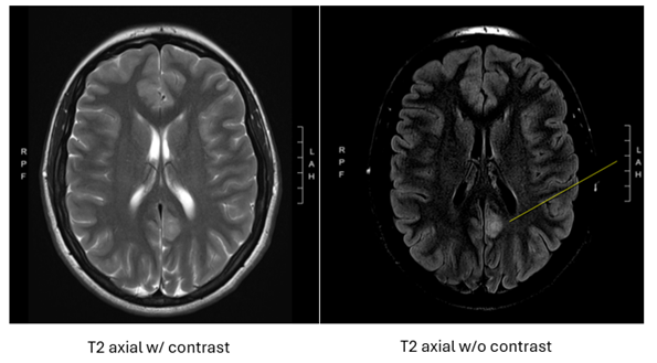
Title: A 25 year old female with new seizure-like activity. Author(s): Heather Ames, MD PhD and Dakota Wheeler, MD Institution: University of Maryland School of Medicine Clinical History: A 25 year old female presents with worsening chronic headaches and vision changes with flashing lights. MRI of the brain showed a 1.6 cm, non-enhancing, T2/FLAIR hyperintense lesion in the left posterior cingulate gyrus, with the radiological differential diagnosis including focal cortical dysplasia and low grade glioma. A stereotactic biopsy was performed. Figure 1: MRI of brain showing 1.6 cm T2 hyperintense lesion.
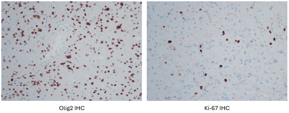
Representative Histology / IHC:
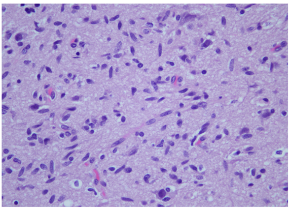
Figure 2: H&E stained slides: Moderately cellular infiltrative glioma with a fibrillary pattern. Perineuronal satellitosis is seen, and there is a delicate branching capillary network in the more cellular portions of the tumor. Tumor cells have ovoid and spindle-shaped nuclei with fine-to-vesicular chromatin and indistinct cytoplasm. There is moderate nuclear atypia, with occasional cells showing marked nuclear pleomorphism. No significant mitotic activity is present and there is no evidence of microvascular proliferation or necrosis. Questions for Viewers: 1) How is the differential diagnosis in this case different than that for a 5 year old or 65 year old? 2) What immunohistochemical stains would help predict the associated molecular changes? 3) How do the potential molecular alterations found in this lesion impact the tumor grade? |
Our Sponsors